Language guided image generation to enhance fracture risk prediction using lateral spine plain radiograph
Authors: Sang Wouk Cho, Yumie Rhee, Kyoung Min Kim, Namki Hong
Journal: ASBMR
Published Date: Nov. 1, 2024
Citation: Cho, Sang Wouk, et al. "Language guided image generation to enhance fracture risk prediction using lateral spine plain radiograph." JOURNAL OF BONE AND MINERAL RESEARCH. Vol. 39. GREAT CLARENDON ST, OXFORD OX2 6DP, ENGLAND: OXFORD UNIV PRESS, 2024.
Abstract:
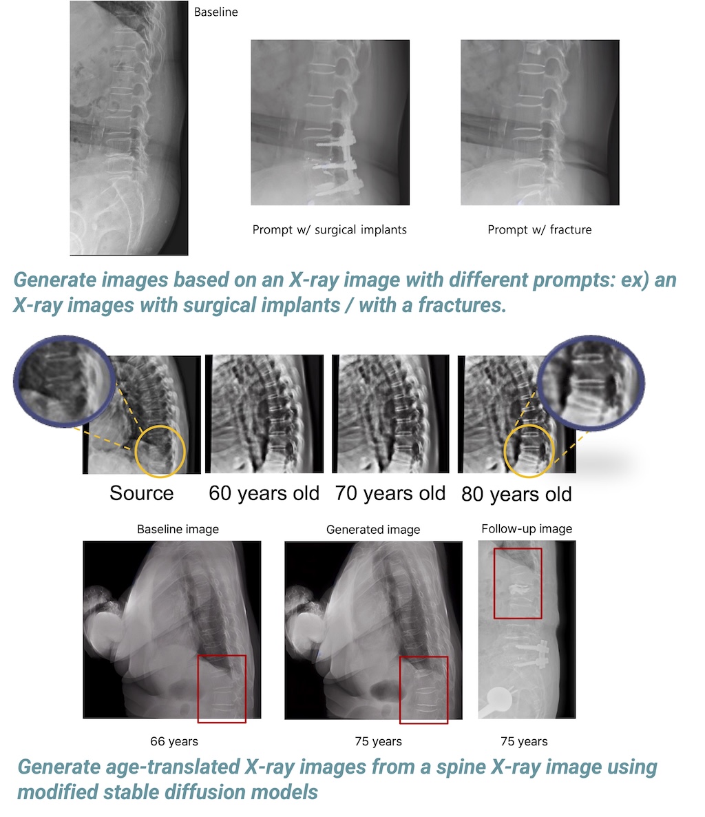
Spine radiography along with deep neural networks is capable of detecting prevalent vertebral fractures and osteoporosis. However, whether the generative model predicts fracture risk remains uninvestigated.
Clinical variables and lateral spine X-ray images from patients aged 50 or older who presented to Severance Hospital, Korea between January 2007 and December 2018 were collected. The incident fracture was defined using follow-up X-ray radiographs. Our model consists of two language-guided latent diffusion models (LDM) to extract feature maps of morphological structure and generate new images with clinical prompts on training set (80% hold-out set) and test set (20% hold-out set). Verte-X prevalent vertebral fracture scores (pVF scores) were calculated on the baseline images (BpVF) and 10-year generative images (GpVF). Fracture risk assessments were then conducted, categorizing them into three groups based on these scores: 1) low risk for both BpVF & GpVF (LL), 2) low risk for BpVF & high risk for GpVF (LH), 3) and high risk for BpVF regardless of GpVF status (HH).
A total of 29,307 lateral spine plain X-rays for 9,276 patients with (mean age 65.7 years, women 66%; VF prevalence 18.6%) were analyzed in the derived cohort. Over a mean follow-up period of 34.8 months, 9.9% of patients experienced vertebral fractures (921 out of 9,276 in the whole dataset) after baseline. Generative images revealed possible changes in the spine at different time points. The mean (SD) error in pVF scores between real-follow up and generative X-ray images was 0.06 ± 0.20 with a correlation coefficient r of 0.655 (0.547,0.741). When stratified into the risk group, LH group and HH risk group were associated with 109% and 391% increased risk of fracture respectively (hazard ratio [HR], 2.092 and 4.911; P=<0.001 for all), showing an improved model fit by adding age, sex, and BMI to covariates (likelihood ratio 105.7, p <0.001). The association between risk groups with incident fracture remained robust ([HR] 1.461; P=<0.001) after adjustment for FRAX major osteoporotic probability.
In summary, generative image-based risk stratification showed its potential to improve clinical workflow and fracture risk assessments.